Imaging
The Park Boush lab recently acquired a Leica M205 C system, which has an optical resolution of 0.952 µm, 20.5:1 zoom, 7.8 x to 160 x magnification and up to 1050 lp/mm resolution (with 2.0x objective). With this imaging capability, comes the ability to do z-stacking and image manipulations.
Macropod Pro
The Macropod Professional & Micro Kit is a lab and field based photomacrography system which uses focus stacking (focal plane merging and z-stacking) to allow for digital image capture in the field down to the ~1 μm scale. It is completely portable and will be taken in the field for in-situ imaging where necessary. This system, combined with a gigapan, can do outcrop level imaging to the grain size of
sand/silt.
Core Viewing Set-Up
The Park Boush Lab has monitors set up in order to look at acquired cores in full view. The ability to stack high quality core photographs allows for detailed analysis of acquired cores without having the physical core present.
Separation Equipment
The Park Boush Lab has separation equipment that includes rock and sediment separators, ultrasonicators, sieves and high precision tools for extracting fossils from rock matrix.
Comparative Collections
Dr. Lisa Park Boush has comparative ostracode material from Lake Tanganyika, the Bahamas, and the Green River Formation.
SEM Facilities
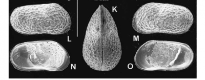
The University of Connecticut has a FEI Nova NanoSEM 450 with low vacuum capabilities to image non- coated specimens such as ostracodes that will be later analyzed for isotopic compositions. The FEI Nova 450 has an ultra-stable, high current Schottky gun. Resolution limits are 1.8 nm @ 3 kV in low vacuum mode 30 Pa. This instrument also includes an Oxford AZtecEnergy Microanalysis System with X-Max 80 Silicon Drift Detector for EDS analyses of shell composition.